|
|
|
Cancer
patients with prickle cell carcinoma of esophagus, malignant melanoma, breast
cancer (4 cases), malignant lymphogranuloma ( 2 cases), gastric carcinoma,
bronchial carcinoid, hepatoma, liver cancer, malignant lymphoma, bladder
cancer, sarcoma, renal cell carcinoma, colon carcinoma, basalioma (2 cases),
carcinoma of the tongue, carcinoma of the prostate, rectal carcinoma,
esophageal cancer, leukemia, melanoma. Control patients with hepatitis,
hyperuricemia, pyelitis, lues, hypertension, psoriasis, chr. tonsillitis,
nephrolittiasis and 5 healthy persons. For the diagnosis and the material I
have to thank R. Pekar.
In the
group of cancer patients we had 90% positive, 6% insecure, 4% negative cultures
(n=48). In the group of patients with other diseases we had 25% positive and
75% negative cultures. In the group of healthy persons we found 20% positive
and 80% negative cultures. We used for the analyses Giemsa stained smears and a
magnification of 1600 : 1 (fig. 4 and fig. 5).
On agar
plates we found two culture types, rough cultures and fried egg cultures (fig.
6a and fig. 6b).
In bouillon
cultivated particles can be seen with dark field microscopy (fig.7). With SEM
we found 0,25µm particles and ring forms (fig. 8 and fig. 9). With the TEM we
found 0,25µm cell-wall-deficient particles (fig. 10) ,0,25µm particles with a
cell wall (fig.10a) and cross sections through ring forms (fig. 11).

Fig.4
Particles (Basoplasma sanguineum),
breast cancer (44a), Giemsa staining, magnification 1600 : 1, positive. (higher resolution)
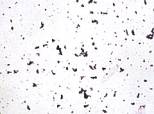
Fig.5
Particles (Basoplasma sanguineum),prickle
cell carcinoma of esophagus, Giemsa staining, magnification 1600 : 1, positive.
(higher resolution)
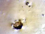
Fig.6a
Colonies of the particles (Basoplasma
sanguineum) on Mycoplasma – agar (Merck) with Mycoplasma-free inactivated
lamb serum. (higher resolution)
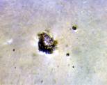
Fig.6b
Colonies of the particles (Basoplasma
sanguineum) on Mycoplasma – agar (Merck) with Mycoplasma-free inactivated
lamb serum.
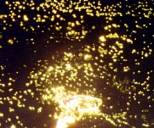
Fig.7
Culture of the particles (Basoplasma
sanguineum) in Mycoplasma – bouillon (Merck) with Mycoplasma-free inactivated
lamb serum, breast cancer, darkfield, magnification 1600 : 1. ( higher resolution)
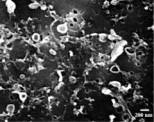
Fig.8
Particles (Basoplasma sanguineum) of
a positive culture (prickle cell carcinoma of esophagus) ,MF- method, REM,
magnification 6250 : 1. (higher resolution)
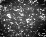
Fig.9
Particles (Basoplasma sanguineum) of
a positive culture (breast cancer) directly smeared on a cover plate, REM,
magnification 6250 : 1 . (higher resolution)
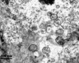
Fig.10 Particles
(Basoplasma sanguineum), cell-
wall-deficient, of a positive culture (prickle cell carcinoma of esophagus),
TEM, magnification 25 000:1. (higher resolution)
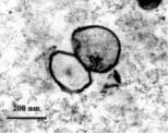
Fig.10a
Particles (Basoplasma sanguineum)
with cell wall, of a positive culture (prickle cell carcinoma of esophagus),
TEM, magnification 25 000:1. (higher resolution)
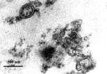
Fig. 11
Particles (Basoplasma sanguineum) of
a positive culture (breast cancer), TEM, magnification 25 000:1. (higher resolution)
Preface Introduction
Blood Analyses Culture
Immunfluorescence Animal
Experiment Discussion Summary Literature Biography