|
|
|
32 mice
Him:OF1 (SD) SFP male. 14 days quarantine; weight 20g , age 6 weeks; diet:
Altromin-R-1324; cage: Makrolon 11
5 groups
A,B,C,D,E à 6 mice
I have to
thank for the animals D. Adamiker and for the histopathological examination W.
Kovac (†).
From the 18
mice of the groups A,B,C with subcutaneous injection of 0,1ml particle
suspension 28% fall ill with cancer (fig.14 to fig. 18a). 39% had chronic
inflammations and 67% had granulocytosis. One mouse from the control had an
adenoma of the lung and one an osteoma that are 17%, the other mice were
healthy.
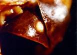
Fig.14
Adenoma of the lung ( 160 days after the beginning of the trial)
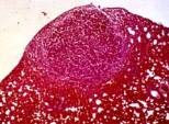
Fig.14a
Adenoma of the lung ( 160 days after the beginning of the trial), H.E. -
staining
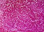
Fig.14b
Adenoma of the lung ( 160 days after the beginning of the trial), H.E. –
staining
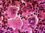
Fig.15
Splenectasis with megakaryocytes ( 160 days after the beginning of the trial),
H.E. – staining
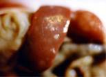
Fig.16
Splenectasis with adenoma ( 379 days after the beginning of the trial)
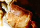
Fig.17
Nephradenoma ( 379 days after the beginning of the trial)
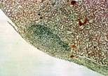
Fig.17a
Nephradenoma ( 379 days after the beginning of the trial), H.E. – staining
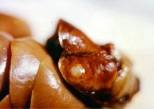
Fig.18
Adenoma of the lung ( 379 days after the beginning of the trial)
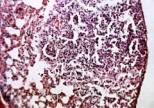
Fig.18a Adenoma
of the lung ( 379 days after the beginning of the trial), H.E. – staining
Preface Introduction
Blood Analyses Culture
Immunfluorescence Animal
Experiment Discussion Summary Literature Biography